AVM Excision / Embolization
Brain / Spine AVM
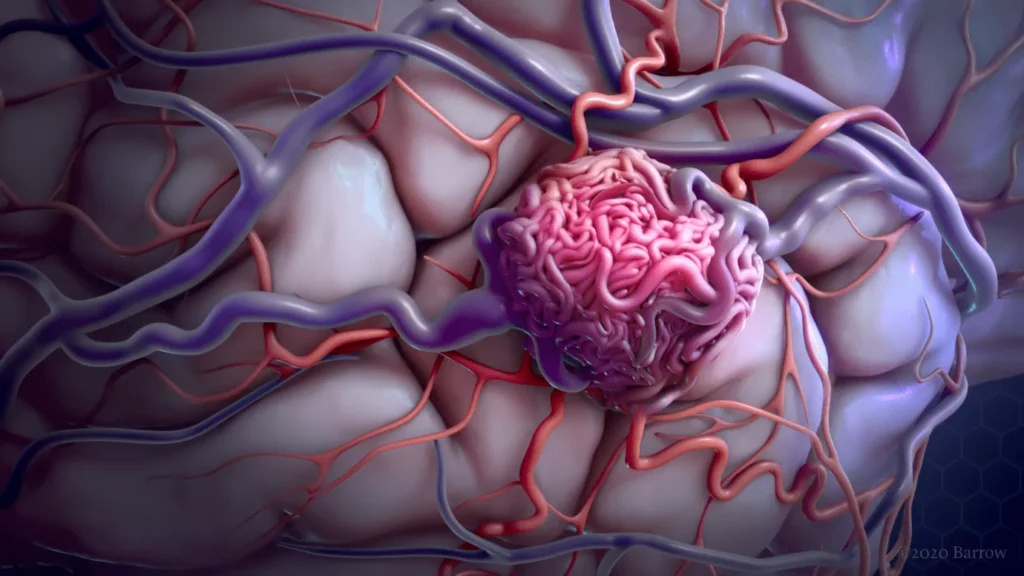
Arteriovenous malformations (AVMs) of the brain or spine refer to abnormal connections between arteries and veins. AVMs can be difficult or dangerous to treat and may cause bleeding into or around the brain, most commonly in young adults. Although AVMs can cause headaches or other symptoms, they are often discovered on CT or MRI scans performed for other reasons.
Left untreated, there is 4 percent risk that they may start to bleed, causing severe neurologic damage and even death.
Although people are born with AVMs, symptoms may not develop until adulthood, often between 20 to 40 years of age.

What Causes Arteriovenous Malformations?
People are born with AVMs, although they do not appear to inherit them from their parents, nor do they give them to their children. It appears that AVMs may be caused by a rupture or clotting of a blood vessel that happens during development before one is born.
What Are the Symptoms of Arteriovenous Malformations?
Seizures and headaches are the most generalized symptoms of AVMs, but no particular type of seizure or headache pattern has been identified. Seizures can be partial or total, involving a loss of control over movement, convulsions or a change in a person’s level of consciousness. Headaches can vary greatly in frequency, duration and intensity, sometimes becoming as severe as migraines. Sometimes a headache consistently affecting one side of the head may be closely linked to the site of an AVM. More frequently, however, the location of the pain is not specific to the lesion and may encompass most of the head.
AVMs also can cause a wide range of more specific neurological symptoms that vary from person to person, depending primarily upon the location of the AVM. Such symptoms may include muscle weakness or paralysis in one part of the body; a loss of coordination (ataxia) that can lead to such problems as gait disturbances; apraxia, or difficulties carrying out tasks that require planning; dizziness; visual disturbances such as a loss of part of the visual field; an inability to control eye movement; papilledema (swelling of a part of the optic nerve known as the optic disc); problems using or understanding language (aphasia); abnormal sensations such as numbness, tingling or spontaneous pain (paresthesia or dysesthesia); memory deficits; and mental confusion, hallucinations or dementia.
What are the treatment options for AVM?
Treatment choices depend on the type, size and location of the AVM, risk of AVM rupture, your symptoms, your age and your general health. Ideally, the goal of treatment is to reduce the chance of bleeding or make it permanently go away. Surgery on your brain and spinal cord is serious, with risks including complications and death. Each person, and each person’s AVM, is unique and there aren’t any perfect decision-making tools in all cases. In general, though, treating an arteriovenous malformation as soon as possible is usually the best way to avoid serious complications.
One or more of these approaches might be tried:
- Surgery to remove the AVM (Excision of AVM).
If the brain AVM has bled or is in an area that can easily be reached, brain surgery to remove the AVM may be recommended. In this procedure, the surgeon removes part of the skull temporarily to gain access to the AVM.
With the help of a high-powered microscope, the surgeon seals off the with special clips and carefully removes it from surrounding brain tissue. The surgeon then reattaches the skull bone and closes the incision in the scalp. Resection is usually done when the AVM can be removed with little risk of hemorrhage or seizures. AVMs that are in deep brain regions carry a higher risk of complications. In these cases, your neurosurgeon provider may recommend other treatments.

- Embolization. In this procedure, a catheter is inserted into an artery in your groin and moved to the location of the AVM. Once there, a glue-like substance, coils or other substance is released into the AVM, which slows or stops the blood flow through the AVM. This approach is used when the AVMs are large and have a lot of blood flow through them.
This way, they can be more easily removed with less risk of bleeding if surgery is performed immediately afterward. Embolization can also slow blood flow to reduce rupture if surgery isn’t immediately performed.
- Gamma knife radiosurgery. This approach uses highly focused beams of radiation that slowly shrink, scar and dissolve an AVM over a few years or make the AVM easier to remove with surgery.
Looking for Neuro
Surgeon?
Simply give us a call and book an appointment for yourself. We are here to help. Walk into our Hospital and let us take a closer look to suggest the best treatment you need.
